

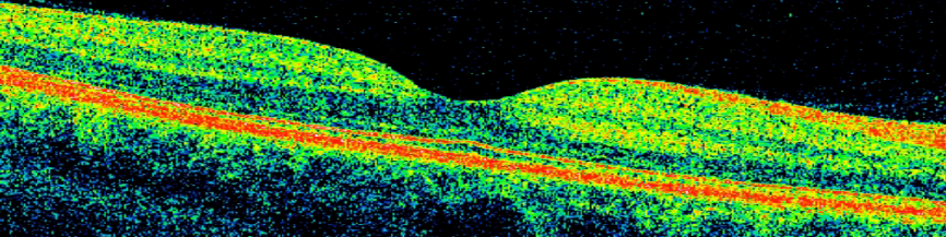
Optical coherence tomography (OCT) macula scan, with color added to highlight retinal neural and supporting layers. Courtesy of Silverstein lab
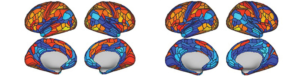
The left panel shows cortical modulations during a visual perceptual organization task; the right panel shows modulations that were predicted from resting-state functional connectivity and brain activity flow mapping (Keane et al., 2021, Neuroimage). Courtesy of Keane lab
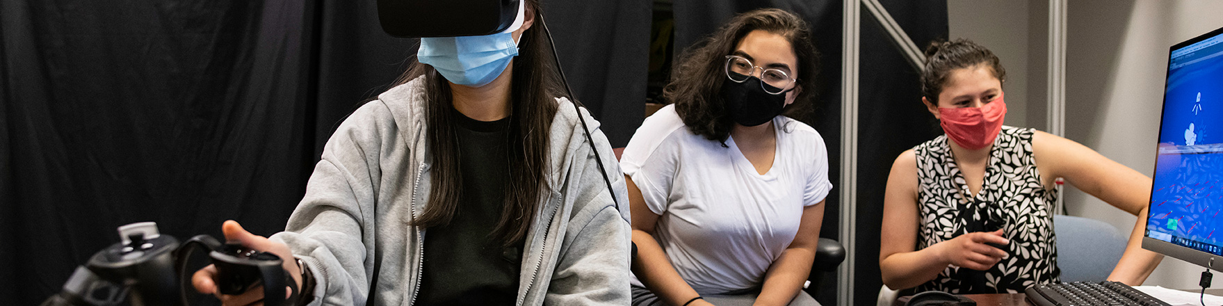
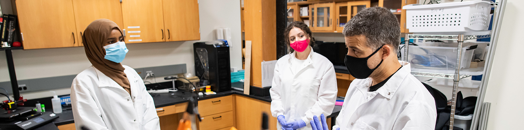
Yacinda Hernandez (C), a senior psychology major at City College New York works with BCS graduate students Ying Lin, left and Emily Isenstein on an experiment accessing visual proprioceptive integration in Meliora Hall. Courtesy of J. Adam Fenster / University of Rochester

OCT macula cube image showing neural layers from the inner limiting membrane (top) to the retinal pigment epithelium (bottom). Courtesy of Silverstein lab
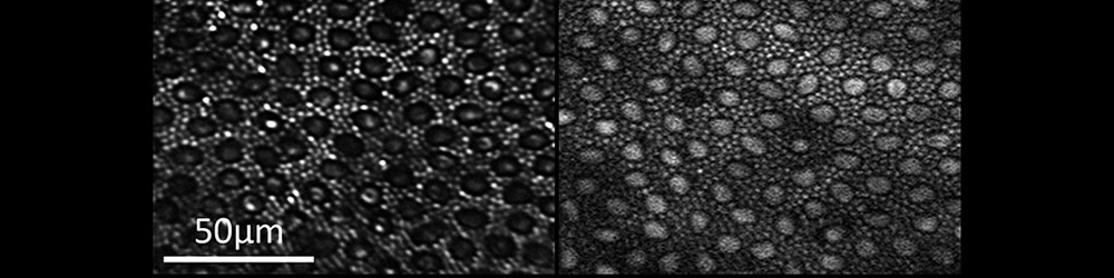
Reflectance (left) and two-photon excited fluorescence (right) image of the photoreceptor mosaic in the living macaque eye. The main source of fluorescence is most likely all-trans-retinol. Courtesy of ARIA
Courtesy of J. Adam Fenster / University of Rochester
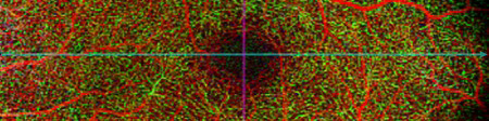
Optical coherence tomography angiography (OCTA) image of the superficial retinal vascular layer, centered on the foveal avascular zone and showing the surrounding microvasculature. Courtesy of Silverstein lab
Ex vivo two-photon microscopy image of ganglion cells below the nerve fiber layer. Courtesy of ARIA
Lulu Abdullahi (L), a junior at East High, practices soldering to repair an experiment component with Manuel Gomez-Ramirez, an Assistant Professor of Brain and Cognitive Sciences, in the Haptics Lab in Meliora Hall. Courtesy of J. Adam Fenster / University of Rochester
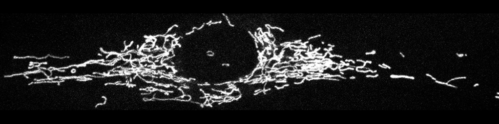
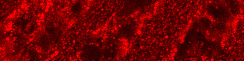
Confocal image of the labeled mitochondrial network inside a corneal fibrobast in culture. Courtesy of Huxlin lab (Ankita Kumar)
Register now for the CVS Symposium
August 15-17, 2024 at the Memorial Art Gallery
Center for Visual Science
The Center for Visual Science (CVS) is an interdepartmental program that brings together vision scientists at the University of Rochester. We are united by a shared conviction that the visual system can only be understood by the coordinated effort of diverse scientists focusing on different parts of the problem.
The expertise of CVS faculty spans psychophysical, optical, physiological, computational, anatomical, and clinical approaches to visual science. The role of the center is to integrate these approaches into a coordinated research effort. Over 40 participating investigators hold their primary appointments in one of eight departments: biomedical engineering, brain and cognitive sciences, computer science, the Flaum Eye Institute, neurology, neuroscience, the Institute of Optics, or Psychiatry, plus the Chester F. Carlson Center for Imaging Science at the Rochester Institute of Technology. We are primarily clustered in two main locations--the School of Medicine and Dentistry and Arts, Sciences, and Engineering. Vision research at Rochester falls within five major themes.