

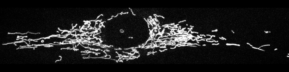
Confocal image of the labeled mitochondrial network inside a corneal fibrobast in culture. Courtesy of Huxlin lab (Ankita Kumar)
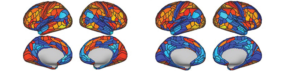
The left panel shows cortical modulations during a visual perceptual organization task; the right panel shows modulations that were predicted from resting-state functional connectivity and brain activity flow mapping (Keane et al., 2021, Neuroimage). Courtesy of Keane lab
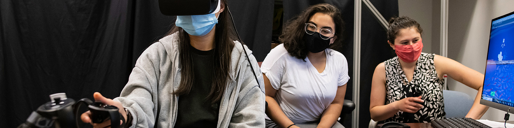
Yacinda Hernandez (C), a senior psychology major at City College New York works with BCS graduate students Ying Lin, left and Emily Isenstein on an experiment accessing visual proprioceptive integration in Meliora Hall. Courtesy of J. Adam Fenster / University of Rochester

OCT macula cube image showing neural layers from the inner limiting membrane (top) to the retinal pigment epithelium (bottom). Courtesy of Silverstein lab
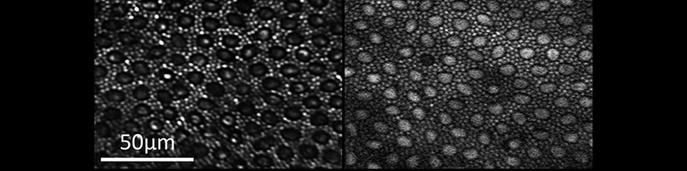
Reflectance (left) and two-photon excited fluorescence (right) image of the photoreceptor mosaic in the living macaque eye. The main source of fluorescence is most likely all-trans-retinol. Courtesy of ARIA
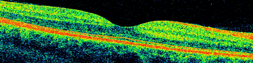
Optical coherence tomography (OCT) macula scan, with color added to highlight retinal neural and supporting layers. Courtesy of Silverstein lab
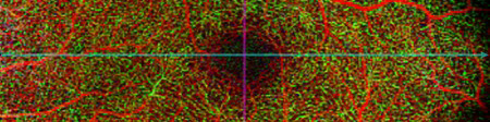
Optical coherence tomography angiography (OCTA) image of the superficial retinal vascular layer, centered on the foveal avascular zone and showing the surrounding microvasculature. Courtesy of Silverstein lab
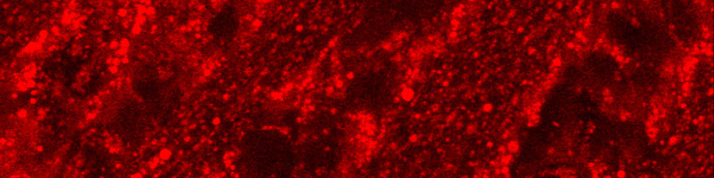
Ex vivo two-photon microscopy image of ganglion cells below the nerve fiber layer. Courtesy of ARIA
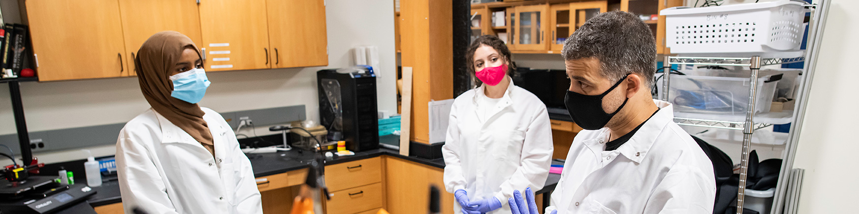
Lulu Abdullahi (L), a junior at East High, practices soldering to repair an experiment component with Manuel Gomez-Ramirez, an Assistant Professor of Brain and Cognitive Sciences, in the Haptics Lab in Meliora Hall. Courtesy of J. Adam Fenster / University of Rochester
Researchers in the Center for Visual Science (CVS) have been at the forefront in developing advanced scientific techniques, including:
- Multi-electrode recordings in awake-behaving monkeys
- Virtual reality tools for studying complex visuomotor behaviors
- Advanced mathematical analysis of behavioral and neural data
- Adaptive optics applications to basic and clinical vision research
- Optical and molecular tools for refractive error correction and vision restoration
CVS had been built on the conviction that progress in vision science requires the coordinated efforts of scientists with very different skills. Researchers in the center apply a number of approaches to their research.
Research Themes at CVS
Visual Perception, Cognition and Action
- Briggs: Understanding vision and attention at the level of neural circuits
- Chapman: Research in brain information processing
- DeAngelis: Neural basis of 3D visual perception and multi-sensory cue integration
- Diaz: Visual guidance of action
- Fiebelkorn: Neural dynamics underlying visual selective attention
- Haefner: Perceptual decision-making
- Huxlin: Characterizing the impact of V1 damage on perception and action
- Jacobs: Visual and multisensory learning and memory, perceptual psychophysics, computational modeling
- Kanan: Brain-inspired and deep neural network models of vision and memory
- Keane: Mechanisms of visual object perception via psychophysics and fMRI
- Lange: Characterizing ‘optimal' visual representations, uncertainty quantification, deep learning
- Marcos: The impacts of optics on visual perception, function and neural adaptation
- Mitchell: Primate visual cortex, active vision, perception, and attention
- Murdoch: Color perception and visual adaptation in advanced displays and lighting systems
- Padmanabhan: Dissecting the neural circuits underlying sensory coding and psychiatric disease
- Poletti: The interplay of vision, eye movements and attention
- Pelz: Eye movements in natural tasks
- Romanski: Functional organization of the primate frontal lobes
- Rucci: Vision and action
- Snyder: Micro- and macro-scale mechanisms of visual attention
- Suarez-Jimenez: Spatial mapping and learning using MRI, VR, and eye-tracking
- Tadin: Mechanisms of visual perception
- Telias: Studying how retinal disease degrades vision and how to restore it
- Zhu: Efficient AR/VR system designs driven by human visual perception
Visual Development, Learning and Plasticity
- Foxe: Basic neurophysiology of schizophrenia and autism
- Huxlin: Using visual learning and plasticity for vision restoration after V1 damage
- Jacobs: Visual and multisensory learning and memory, perceptual psychophysics, computational modeling
- Kanan: Brain-inspired and deep neural network models of vision and memory
- Majewska: Imaging synaptic structure and function in the visual system
- Padmanabhan: Dissecting the neural circuits underlying sensory coding and psychiatric disease
- Suarez-Jimenez: Spatial mapping and learning using MRI, VR, and eye-tracking
- Tadin: Perceptual learning and cognitive training
- Telias: Maladaptive plasticity of retinal neurons in blindness
Multisensory and Sensorimotor Integration
- DeAngelis: Neural basis of 3D visual perception and multi-sensory cue integration
- Gomez-Ramirez: Neural dynamics in cross-modal circuits mediating haptics
- Jacobs: Visual and multisensory learning and memory, perceptual psychophysics, computational modeling
- Lalor: Modeling the neurophysiological processing of natural stimuli in humans
- Poletti/Rucci: Retinal, motor, and proprioceptive integration in the establishment of visual representations
- Romanski: Functional organization of the primate frontal lobes
- Schieber: Neural control of hand and finger movements
Advanced Optical Technology
- Fienup: Image processing, wavefront sensing
- Gomez-Ramirez: Optogenetics stimulation and calcium imaging in mammalian brains
- Kanan: Medical computer vision
- Knox: Femtosecond laser technology for vision
- Marcos: Ocular wavefront sensing, adaptive optics vision simulator and high-resolution anterior segment imaging
- McGregor: Optogenetic vision restoration and the physiology of the fovea
- Merigan: In vivo adaptive optics imaging of the retina
- Poletti/Rucci: New methods for eye-tracking, gaze-contingent display control, virtual and augmented reality
- Rolland: Optical system design and instrumentation for imaging science and 3D visualization
- Schallek: Imaging blood flow in the living eye
- Williams: Limits of human vision
- Zavislan: Optical system design for clinical diagnostics
- Zheleznyak: Femtosecond laser technology for vision and optical metrology
Disorders of Vision
- Buckley: Soft tissue biomechanics
- DiLoreto: VEGF trap treatment for age related macular degeneration, age related eye disease, adaptive optics imaging of inherited macular diseases
- Feldon: Orbital disease and neuro-ophthalmology
- Foxe: Basic neurophysiology of schizophrenia and autism
- Huxlin: Perceptual and molecular approaches to vision restoration
- Keane: Visual differences in psychosis and their clinical significance
- Levin: Understanding genotype-phenotype correlations and developing gene based and gene agnostic therapeutics for inherited retinal disorders
- Libby: Neurobiology of glaucoma
- MacRae: Refractive surgery
- Marcos: Optical vision correction
- McGregor: Optogenetic vision restoration and the physiology of the fovea
- Merigan: In vivo adaptive optics imaging of the retina
- Poletti/Rucci: Impairments in eye movements, attentional control, and active vision
- Schallek: Imaging blood flow in the living eye
- Silverstein: Retinal biomarkers of brain structure and function in schizophrenia and other neuropsychiatric disorders
- Singh: Cellular and molecular mechanisms of retinal and neurodegenerative diseases
- Tadin: Vision in special populations
- Telias: Retinitis pigmentosa, age-related macular degeneration, photoablation
- Woeller: Understanding the key molecular and cellular pathways involved in eye disease, with a particular focus on Thyroid Eye Disease (TED)
- Wozniak: Understanding how and why bacterial infections cause ocular tissue damage, as well as the development of novel ophthalmic antimicrobial therapeutics to treat this blinding disease
- Zheleznyak: Novel optical therapies to treat myopia progression, presbyopia and refractive error
- Zhu: Computational and assistive technologies to treat color vision deficiency